Abstract
Linear IgA bullous disease (LABD) is a rare autoimmune blistering disorder characterized by the linear deposition of IgA antibodies along the basement membrane zone. This article provides a comprehensive overview of LABD, covering demographics, causes, clinical features, complications, diagnosis, differential diagnoses, treatment modalities, and outcomes, aiming to enhance understanding and facilitate improved management of this unique dermatological condition.
1. Introduction
Linear IgA bullous disease (LABD) is an autoimmune blistering disorder characterized by the linear deposition of IgA antibodies along the basement membrane zone. This condition results in the formation of blisters and erosions on the skin and mucous membranes, posing diagnostic challenges due to its diverse clinical manifestations.
2. Demographics
Age of Onset: LABD can affect individuals of all ages, but it commonly presents in childhood or adulthood.
Gender: There is no predilection for gender, with LABD affecting both males and females.
Incidence: LABD is considered rare, with an estimated incidence of 0.5 to 2 cases per million population per year.
3. Causes
- Autoimmune Nature: LABD is an autoimmune disorder where the immune system mistakenly produces antibodies against components of the basement membrane zone, particularly the IgA class.
- Triggers: Various factors, including infections, medications, and underlying inflammatory conditions, may trigger LABD in genetically predisposed individuals.
4. Clinical Features
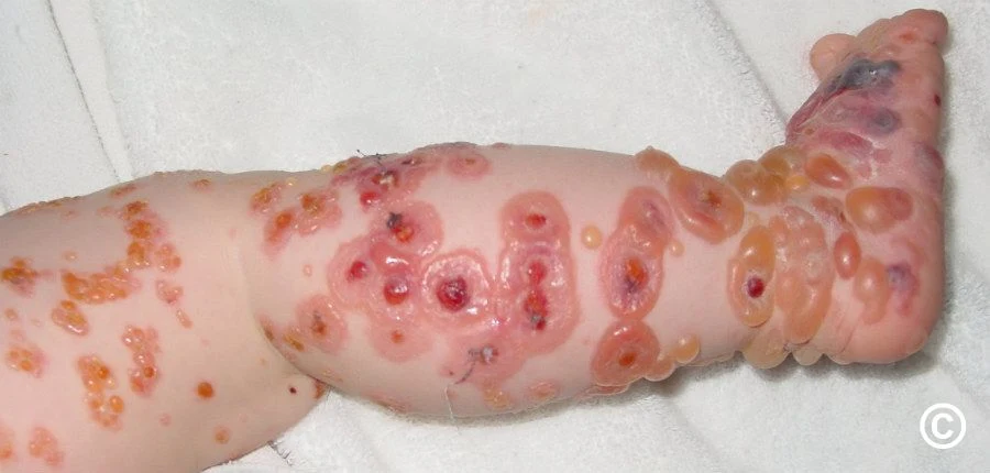
- Lesion Distribution: LABD typically presents with tense blisters and erosions affecting the skin, particularly in flexural areas. Mucous membrane involvement is common.
- Nikolsky Sign: Positive Nikolsky sign, where slight rubbing of the skin leads to the formation of new blisters, is often observed.
- Variants: LABD can manifest in different clinical variants, including a childhood variant known as chronic bullous disease of childhood.
5. Complications
- Secondary Infections: Open blisters and erosions can predispose individuals to secondary bacterial infections.
- Scarring: Prolonged and severe cases of LABD may result in scarring and pigmentary changes.
6. Diagnosis
- Histopathology: A skin biopsy, particularly a perilesional biopsy, is essential for diagnosis. Histopathological examination reveals subepidermal blistering.
- Direct Immunofluorescence: The linear deposition of IgA along the basement membrane is a hallmark finding in direct immunofluorescence studies.
7. Differential Diagnoses
LABD needs to be differentiated from other autoimmune blistering disorders, including bullous pemphigoid, dermatitis herpetiformis, and epidermolysis bullosa acquisita.
8. Treatment
- Systemic Corticosteroids: Oral corticosteroids are often used as the first-line treatment to control disease activity.
- Dapsone: Dapsone, an anti-inflammatory medication, is commonly employed in the management of LABD.
- Immunosuppressive Agents: In severe cases or when steroid-sparing agents are needed, immunosuppressive drugs like azathioprine may be considered.
9. Outcome
The prognosis of LABD varies, with some cases experiencing a chronic and relapsing course, while others may achieve remission with appropriate treatment. Regular follow-up is essential to monitor disease activity and adjust treatment as needed.
Conclusion
Linear IgA bullous disease is a complex autoimmune blistering disorder that necessitates a multidisciplinary approach for accurate diagnosis and optimal management. Ongoing research into the pathogenesis of LABD may offer novel insights and therapeutic strategies to improve outcomes for affected individuals.
References:
1. Fortuna G, Marinkovich MP. Linear immunoglobulin A bullous dermatosis. Clin Dermatol. 2012;30(1):38-50.
2. Wojnarowska F, Marsden RA, Bhogal B, Black MM. Chronic bullous disease of childhood, childhood cicatricial pemphigoid, and linear IgA disease of adults. A comparative study demonstrating clinical and immunopathologic overlap. J Am Acad Dermatol. 1988;19(5 Pt 1):792-805.
3. Murrell DF, Daniel BS, Joly P, et al. Definitions and outcome measures for mucous membrane pemphigoid: recommendations of an international panel of experts. J Am Acad Dermatol. 2015;72(1):168-174.


Post a Comment
Full Name :
Adress:
Contact :
Comment: